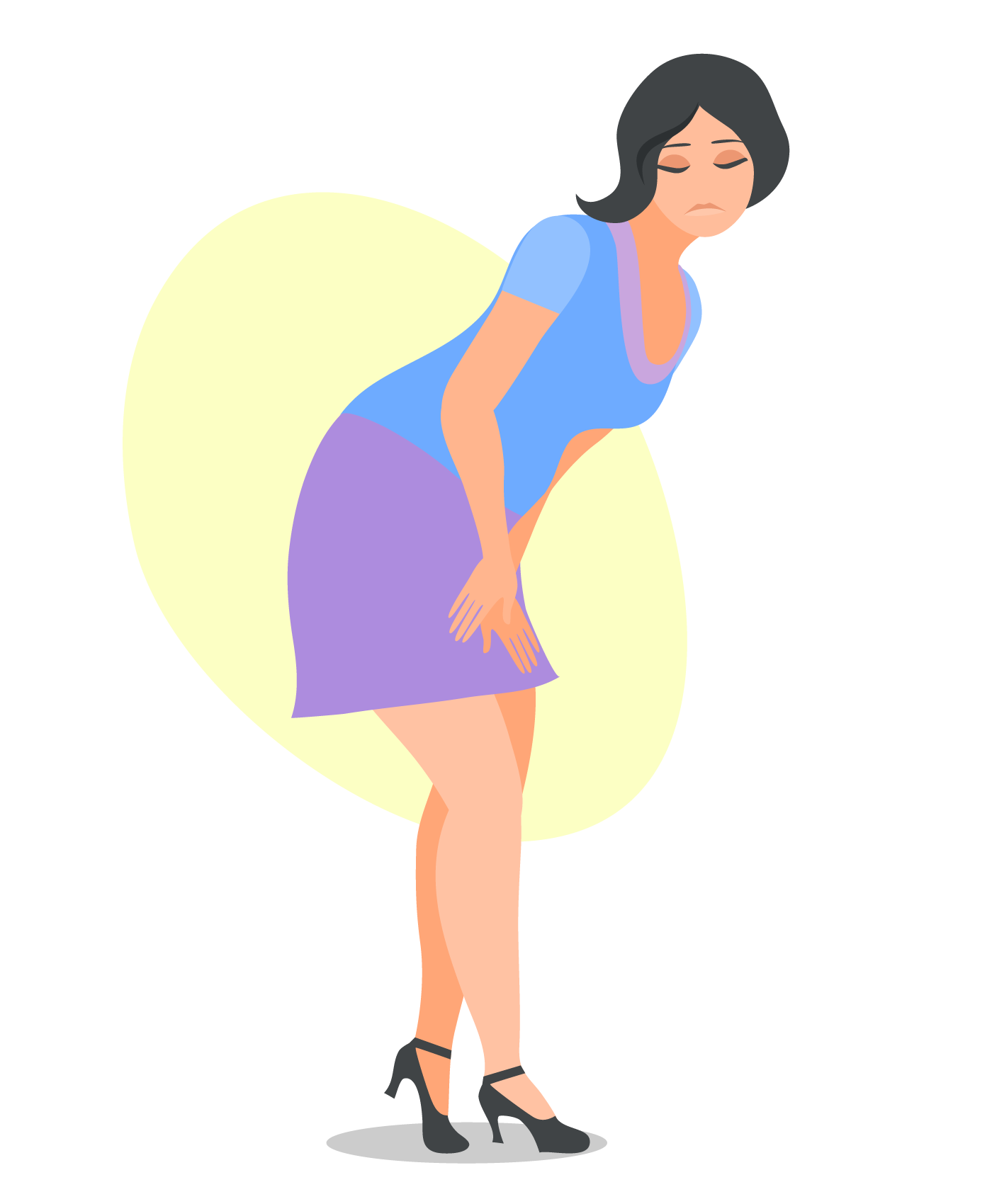
Definition –
Involuntary leakage of urine that is sufficient enough in frequency and amount to cause physical and/or emotional distress.
Frequency –
Highly prevalent in women across their adult lifespan and severity increases linearly with age in women across their adult lifespan: 4–8% ultimately seek medical attention. One in three women aged >60 years has a bladder control problem.
Cause –
Continence and urination involve a balance between urethral closure and detrusor (bladder smooth muscle) activity. Urethral pressure normally exceeds bladder pressure, resulting in urine remaining in the bladder. Intra-abdominal pressure increases (coughing, sneezing) are normally transmitted to both urethra and bladder equally, maintaining continence. Disruption of this balance leads to various types of incontinence.
Diagnosis-
1 History –
A detailed history is important to determine the severity of symptoms and rule out medication causes. Emotional distress often does not correlate well with the amount of urine loss that can be demonstrated.
2 Physical examination (three areas)-
• General examination to rule out delirium and, atrophic urethritis, restricted mobility, or stool impaction.
• Urogynecologic examination may reveal severe vulvar excoriation from continual dampness. The vaginal tissue should be inspected for signs of atrophy, stenosis, bladder neck mobility (Q-tip test), and atrophic urethritis. The patient is asked to cough repeatedly or undergo a Valsalva maneuver with a full bladder in the lithotomy or standing position to induce urine leakage. Rectal examination can evaluate rectal sphincter tone or the presence of fecal impaction.
3 Urinalysis and urine culture –
Many relevant metabolic and urinary tract disorders can be screened by a simple urinalysis. A culture is essential to rule out infection before proceeding with further evaluation.
4 Residual urine volume after voiding –
A catheterized post-void residual (PVR) urine specimen should be obtained to exclude urinary retention (normal PVR ≤100 mL) or infection.
5 Frequency –
Volume bladder chart. More than seven voids per day suggest a problem with frequency, but this is highly dependent on habit and fluid intake. Patients can be notoriously inaccurate in estimating urinary frequency and should be encouraged to keep a “urinary diary” for several days as part of their initial evaluation.
6 Urodynamics –
It is a group of tests designed to aid in determining the etiology of lower urinary tract dysfunction.
• Simple cystometry involves placing a catheter and gradually filling the bladder with sterile water. Involuntary “detrusor” contractions are demonstrated by a rise in water level during filling due to back-pressure. Normally, the first sensation to void occurs at 150 mL and bladder capacity is typically 400–600 mL.
• Uroflowmetry is used to determine the urinary flow rate and flow time to screen for the presence of outflow obstruction and abnormal detrusor contractility. Normally, women achieve a peak flow rate of 15–20 mL/s with a voided volume of 150–200 mL.
• Complex urodynamic testing requires placement of an intravesical catheter to measure detrusor pressures and a vaginal or rectal catheter to indirectly measure intra-abdominal pressures.
Stress urinary incontinence (SUI)
Patients have loss of small amounts of urine with coughing, laughing, sneezing, exercising, or other movements that increase intra-abdominal pressure and thus increase pressure on the bladder.
• Etiology –
Physical changes resulting from pregnancy, childbirth, and menopause often result in weaknesses in the pelvic floor, and urethral support structures and nerve damage.
• Mechanism –
If the fascial support is weakened, the urethra can move downward at times of increased abdominal pressure, causing bladder pressure to exceed urethral sphincter closure pressure (hypermobile urethra). Incomplete urethral closure may be due to scarring or neuromuscular damage, and cause a more severe form of stress urinary incontinence – intrinsic sphincter deficiency.
• Diagnosis –
SUI is suggested by history, physical examination, and a positive stress test (demonstrable loss of urine while the patient is being examined).
• Non-surgical treatment –
Pelvic muscle (Kegel) exercises, biofeedback (pressure measurement device notifies the patient when correct muscle contraction is performed and reinforces correct technique), pessaries.
• Surgical treatment :
1 Tension-free transvaginal tape (TVT) or trans obturator tape (TOT) are minimally invasive suburethral sling procedures that are rapidly becoming the “gold standard.”
2 Burch colposuspension involves suture placement at the Cooper ligament. The Marshall–Marchetti–Krantz (MMK) variation has sutures going through the periosteum of the pubic symphysis.
3 Anterior colporrhaphy has a poor long-term success rate.
4 Collagen periurethral injections (Coaptite, Macroplastique) are designed as a treatment for SUI resulting from intrinsic sphincter deficiency.
Urge incontinence
Patients experience involuntary leakage for no apparent reason while suddenly feeling an urgent need to urinate. This may be accompanied by urinary frequency and nocturia, and patients often describe their bladder as “spastic” or “overactive.”
• Etiology –
Involuntary detrusor muscle contractions. Detrusor hyperactivity can be due to loss of central nervous system (CNS) inhibitory pathways, local irritants, or bladder outlet obstruction.
• Mechanism –
Frequently idiopathic, but results from damage to the nerves of the bladder, the nervous system (spinal cord and brain), or the muscles themselves.
• Treatment –
Behavior modification (bladder drills, biofeedback) and/or pharmacologic therapy (oxybutynin chloride, imipramine, mirabegron), injection of the detrusor muscle with botulinum toxin A, neuromodulation.
Overflow incontinence
Patients experience continuous, unstoppable dribbling of urine, or continuing to dribble for some time after they have passed urine.
• Etiology –
The bladder is always full and overflows, resulting in frequent or continuous urine leakage.
• Mechanism –
Weak bladder detrusor muscles, resulting in incomplete emptying, or a blocked urethra (outflow obstruction) due to advanced vaginal prolapse, or after an anti-incontinence procedure that has overcorrected the problem.
• Treatment –
Catheter drainage, followed by treatment of the underlying condition.
Other types of incontinence
• Mixed incontinence-
It usually refers to the common combination of stress and urge incontinence occurring together.
• Transient incontinence –
It is often triggered by medications, urinary tract infections, mental impairment, restricted mobility, or stool impaction (severe constipation), which can push against the urinary tract and obstruct outflow.
• Functional incontinence –
It occurs when a person does not recognize the need to go to the toilet, recognize where the toilet is, or get to the toilet in time due to confusion, dementia, poor eyesight, or poor mobility.